


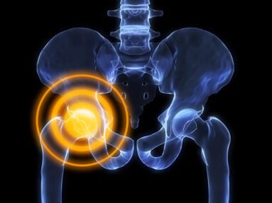
Hip arthrosis is a complex disease with specific symptoms and complex treatment.The disease occurs against the background of metabolic disorders in the cartilage tissue of the joint cavity and the head of the femur.
Hip arthrosis or coxarthrosis is more common in the elderly.It is generally accepted that the inflammatory reaction plays the main role in the pathogenesis of the disease.After a large number of studies, arthrosis has been shown to occur with atherosclerosis and diseases with metabolic disorders.
The essence of the disease
Coxarthrosis is a disease based on metabolic disorders with atrophic and degenerative changes in the cartilage tissue of the hip joint.
You cannot confuse arthritis with arthritis.Unlike arthritis with arthrosis, a non -infectious (aseptic) inflammation occurs, which develops and progresses for many years.
Pathogenesis of Development:
- Disruption of metabolic processes in cartilage.Cartilage tissue gets nutrients using diffusion.The slightest inflammation or edema leads to a lack of trace elements and minerals.
- Against the background of processes impaired by food, atrophic changes begin, cartilage tissue is refined, the amount of joint fluid and chondroblasts decreases.
- Due to the madness and destruction of cartilage, severe pain begins, there is a decrease in the amplitude of movements in the joints.
- The material of the cartilage is very sophisticated, the fabulous cleft narrows, becomes joint dystrophy.
Dystrophy for more than one year, more than one year, has been held.The disease can only be discontinued in the first stages, with the development of the third stage of osteoarthrosis, treatment is aimed at reducing the symptoms and claiming the patient's life, an alternative to medicines - endoprothetics.
Reasons
The disease is polyetiological, there are many conditions and factors that can lead to arthrosis or provoke its progression.If the causes of hip arthrosis are not detected, such a disease is called idiopathic arthrosis.
The disease is not hereditary, but genetic pathologies in which cartilage dysplasia occurs can cause arthrosis of the hip joint.
Also, the cause of coxarthrosis can be such diseases:
- Pertteres Syndrome - The characteristic sign of the disease is a disorder of the supply of nutrients to the cartilage tissue of the joint and head of the femur.This happens in childhood, mainly the boys are sick.
- Congenital dislocations and subluxation of the femur.In the process of injury, an inflammatory reaction and aseptic melting of the cartilage and the head of the femur may occur.
- Necrosis of the femur head.It occurs due to damage to the upper artery, which is attached to the upper part of the head.
- Rheumatoid and youthful arthritis.Against the background of the action of toxins or own antibodies, exudative inflammation in the joint develops.
Given the fact that the disease progresses slowly, the disease can be from one and two.
There are many factors contributing to the onset of arthrosis, they include:
- Spinal diseases (kyphosis, lordosis, scoliosis);
- metabolic diseases of connective tissue;
- violation of the blood supply to the joint;
- atherosclerosis of large vessels;
- states of stress;
- Hip dysplasia;
- congenital deformities of the lower limbs;
- infectious diseases;
- inactive lifestyles;
- alcohol intake, smoking;
- Old age.
Remember that people involved in stretching are at high risk of developing arthrosis in adulthood.
Also, one of the reasons may be traumatic damage to the components of the joint.After the onset of tissue damage, an inflammatory reaction occurs, resulting in the cartilage can be replaced by a clutch.
Symptoms
Due to the fact that the disease is slowly progressing, the patient does not always pay attention to his first signs.It should be noted that with early diagnosis, the chances of remission of the disease increase.It is very important to start treatment early, as it is possible to avoid the onset of ankylosis and complete osteoarthrosis.
With arthrosis of the hip joint, symptoms of varying intensity may occur depending on the workload and the degree of the disease.
Clinical picture of osteoarthritis of the hip joint:
- Painful sensations that develop in severe front pain and lateral thigh.Patients complain that the thigh hurts significantly during the bend or load on the joint.
- The unpleasant sensations arising in the groin when walking, sometimes they are combined with thigh pain.
- The connectivity and restriction of the mobility of the limb in the hip joint.First, the function of leaving aside suffers, and then everyone else.
- Unpleasant sounds when walking, the joint can click or crush.Constant pathological sounds can be the only sign of the disease.
- Morning stiffness that passes after a few hours or before dinner.
Sometimes ignoring the possible consequences, people begin to take medicines for symptomatic therapy and thus mask the progression of the process of destruction in cartilage.
Disease
The clinical picture depends on the degree of arthrosis of the hip joint and the reactivity of the patient's body.If symptoms occur, as a rule, changes occur in the X.In medical practice, it is customary to distinguish three radiological stages, each of which has its own characteristics.
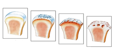
The degree of arthrosis in terms of changes in the X:
1st degree arthrosis
It continues with minimal clinical manifestations and therefore patients rarely seek help from a doctor.With early diagnosis of the disease, the patient increases the chances of full recovery.The initial period of the disease is characterized by small pelvic and thigh pain, the pain increases in the background of exercise or long walking.Secondly, in terms of the frequency of manifestation comes a symptom of groin pain.At 1 degree, the pain is pulled and rarely occurs.The volume of movements is completely preserved.In X -ray, minor changes are displayed.
2nd degree
In the case of a second degree, the patient begins to worry more and more pain that can occur at rest.Symptoms, as a rule, manifest themselves in the evening and morning stiffness does not go to dinner.During prolonged transitions, a symptom of the lame arises, one cannot completely load the patient.The discomfort occurs during flexion or squat, degenerative processes progress in the cartilage.Against the background of such changes, the foot may be shortened, atrophy of the hip muscles and the pelvis appears.Radiographs show the narrowing of the joint gap, the periosteal reaction increases.A large number of osteophytes were found in the lumen of the joint.
The final or diffuse 3rd stage of arthrosis
The third stage is characterized by the appearance of motor dysfunction of the lower limbs.The patient complains of constant pain that happens for no reason.There is a shortening of the limb over 5%, ankylosis occurs, the joint loses the ability to mobility.Radiography shows the complete closure of the joint gap and a large number of osteophytes against the background of bone deformity.Grade 3 treatment is performed only in an operational manner.
Methods of treatment
The choice of treatment methodology depends on the degree of osteoarthritis.In the first stages, complete conservative treatment is used.The most difficult is the second stage, as conservative therapy is ineffective and indications of surgery are not sufficient.It is quite possible to cure arthrosis only with the development of the first degree of the disease.
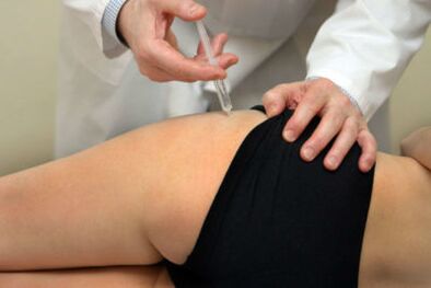
After diagnosing arthrosis of the hip joint, the doctor chooses treatment methods.Most often used:
- conservative treatment with drugs;
- surgical treatment;
- Exercise and massage therapy.
Each of the treatment methods has its own characteristics, variations and specific goals.Conservative therapy is used for such purposes:
- Fighting the etiological factor.For example, metabolic or hormonal disorders can be corrected.
- Symptomatic treatment aimed at alleviating the patient's life and relieving the symptoms of alaration.Non -steroidal anti -inflammatory drugs are used for this purpose.Most often sodium diclofenac, nimesulide, ibuprofen are used by NSAIDs.
To get rid of constant pain, NSAIDs are taken almost every day and this can affect the patient's gastrointestinal tract and cause the development of peptic ulcer.
Surgical intervention is shown with the third degree of arthrosis and is the only method of restoring walking function.The essence of the methodology is the complete or partial replacement of the joints of the joint with titanium endoprosthesis.
Media physical education is an integral part of all rehabilitation measures.Exercise and massage therapy are aimed at improving blood flow to the joint.Also, exercise therapy is used to reduce the risk of ankylosis.
You should be careful when performing exercises as you can damage the joints osteophytes.Tactics and exercises should be selected by a doctor based on your individual characteristics and clinical picture of the disease.